Breast Thermography
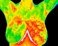
The below image is a normal breast study, with no significant thermal asymmetries.
")
Thermography, also known as, Digital Infrared Thermographic Imaging (DITI) is a safe, non-invasive, painless, radiation free screening test that can detect the subtle physiologic changes in breast tissue that are a result of breast disease. It has the capability to detect the early danger signs of breast cancer, fibrocystic disease, infection or vascular diseases years earlier than is detectable by self breast exam, doctor exam, ultrasound or mammography alone. It may even detect abnormalities in the breast 5-10 years before a mammogram may detect it.
It is based on the principle that in pre-cancerous and early cancerous tissues, metabolic activity and circulation are both increased as compared to normal tissue. When cancer cells implant within the body, they give off many chemicals which initiate the formation of new blood vessel formation around where the cancer cells are multiplying. This process is called neoangiogenesis and is some of the earliest signs that cancer may be developing. The thermography camera will pick up the sympathetic response from this on the dermal circulation which is about 5 mm deep under the skin surface.
How specifically does Thermography detect alterations in blood flow?
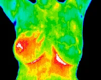
The Thermography camera detects various temperature grades within the body. This is called “thermal imaging”. The images (pictures) will show up in various colors depending upon the amount of warmth or coolness in that particular area. For example, in areas of the body that are generating a lot of heat, the image will show up red or white. In contrast, in areas of the body that are cool or cold, the image will show up as blue or black. Other colors in between hot and cold may show up yellow or green. You will be able to see your own thermal images once you receive a copy of your report.
What is connection between the various colors of the Thermography thermal images and early abnormal tissue changes?
Certain areas of the body are normally at certain temperatures. When variations occur within these normal temperatures, then it can be a “red flag” that something abnormal is developing.
The Thermography camera has the ability to detect alterations in temperatures. For example, the breast tissue is normally a cooler area of the body and normally shows up within the cooler ranges of black or blue with Thermography imaging. Early in the cancer, when the process of neoangiogenesis (new blood vessel formation) is occurring, there will be any increase in heat due to fact that where there is an increase in blood flow, Therefore, the Thermography image would show up in this area as white or red.
What other things could Breast Thermography detect? Thermography can also detect:
|
Fibrocystic Disease |
|
| Infection (ex. mastitis) |
|
|
Vascular Disease |
|
|
Inflammatory Breast Cancer – which is not detected by mammogram |
|
How is Thermography different than Mammography? Thermography is a totally different test as compared to mammography. Thermography has the ability to detect pre-cancerous and early cancerous tissue changes much earlier than mammography, sometimes up to 5-10 years earlier detection.
Mammography looks for “structural changes.” This means that there has to be an actual approximately pea-sized tumor present before the mammography x-ray can see it. On average, it takes about 5-10 years of abnormal cell division before the tumor is pea-sized and therefore large enough for mammography to detect it.
Thermography looks for “physiological changes.” This means that it looks for the early changes that occur years before the tumor is present. This is why Thermography may detect cancer for up to 10 years before the mammogram. And early detection is the key to being able to possibly reverse these abnormal changes before they are a deep-seeded cancer.
Is Thermography approved by the FDA? Yes, thermography is approved by the FDA for breast screening.
Is it okay to do both Thermography and Mammography? Yes, they are completely separate screening tests. It is best to do both. We would never tell you that thermography should replace mammograms but thermography is a valuable adjunct in the evaluation of breast health.
How is the procedure done?
Thermography is a very simple, private and easy procedure. You will be taken to a private imaging room. The technician who will be performing your procedure may ask you a few questions about your paperwork before the test. The entire procedure will be explained to you in advance. The technician will instruct you that she will be leaving the room so that you may get ready for the procedure. You will remove any jewelry from around your neck and earrings. If your hair is long or touches the neckline, you will need to put up your hair so that it does not obscure the neckline in the images. You will then remove your shirt and bra for the screening test and sit on a stool in front of the Thermography camera. The technician will dim the lights for your privacy and re-enter the room where the images will be done at different positions while you sit on the stool throughout the entire process. Once the screening is complete, the technician will leave the room so that you can re-dress. The entire process should not take more than about 10 minutes.
Will I need to return for any further Thermography scans? Yes. After your initial Thermography screening, you will need to return for a follow-up test in approximately 3 months to establish what is called “your baseline pattern”. The reason for this is that vascular patterns (blood vessels) can vary from individual to individual, similar to the way fingerprints vary from person to person. Normally, your vascular patterns do not change unless there is something abnormal occurring. And abnormal changes can be seen with Thermography within 3 months. Basically, you want the follow-up screening to look virtually identical to the first scan. This gives us what this is your normal pattern. Once the baseline is established, then Thermography is recommended annually. All future Thermography scans will be compared to the baseline pattern, as well as, any other previous scans. This helps the doctors to determine if there are any detectible abnormal tissue changes occurring very early in their development.
Is Thermography safe? Yes, Thermography is 100% safe. There is no radiation or any harm to the body whatsoever with the procedure. Thermography may be done at any age and on anyone with absolutely no harm to anyone.
Is there any pain with Thermography? No, no pain at all. It is also non-invasive. In fact, nothing touches your body at all. There is no compression of the breast so your breasts do not get “squished” like with mammograms.
Can Thermography be used to monitor any other things? Yes, it is great for monitoring:
During the course of cancer treatments
Young people with genetic markers/or a strong history of breast cancer
Those with breast implants
Fibrocystic breasts
Large, dense breasts
Identifying lumps without biopsy
Women who decline mammograms
Frequent monitoring without radiation
Is there anything I need to do to prepare for my Thermography? Yes. It is best to not wear deodorant or antiperspirant to the procedure. Also, no heavy exercise prior to the scan.
Is Thermography covered by my health insurance? No, Thermography screening is not covered by health insurance.
Is it expensive? No, Thermography is quite inexpensive as compared to the cost of other diagnostic tests.
Who evaluates the Thermography images? The images are sent via a secure medical server to be read by Medical Doctors who are certified in Thermography reading. The report which includes your images is sent back to our office. Once we receive your report, our doctors will evaluate it and call you if there are concerns. We will also mail a copy of your report with your images to you.
What if I have questions about my report? You may call our office to speak with one of our doctors if you have any questions.
Can Thermography be done on other regions of the body? Yes. Thermography has many other uses other than just for Breast Health screening. Please click on the link below to read about other uses for Thermography.
(link)
Ready to schedule your appointment now? Click on the link or call our office @ 770-719-8785.